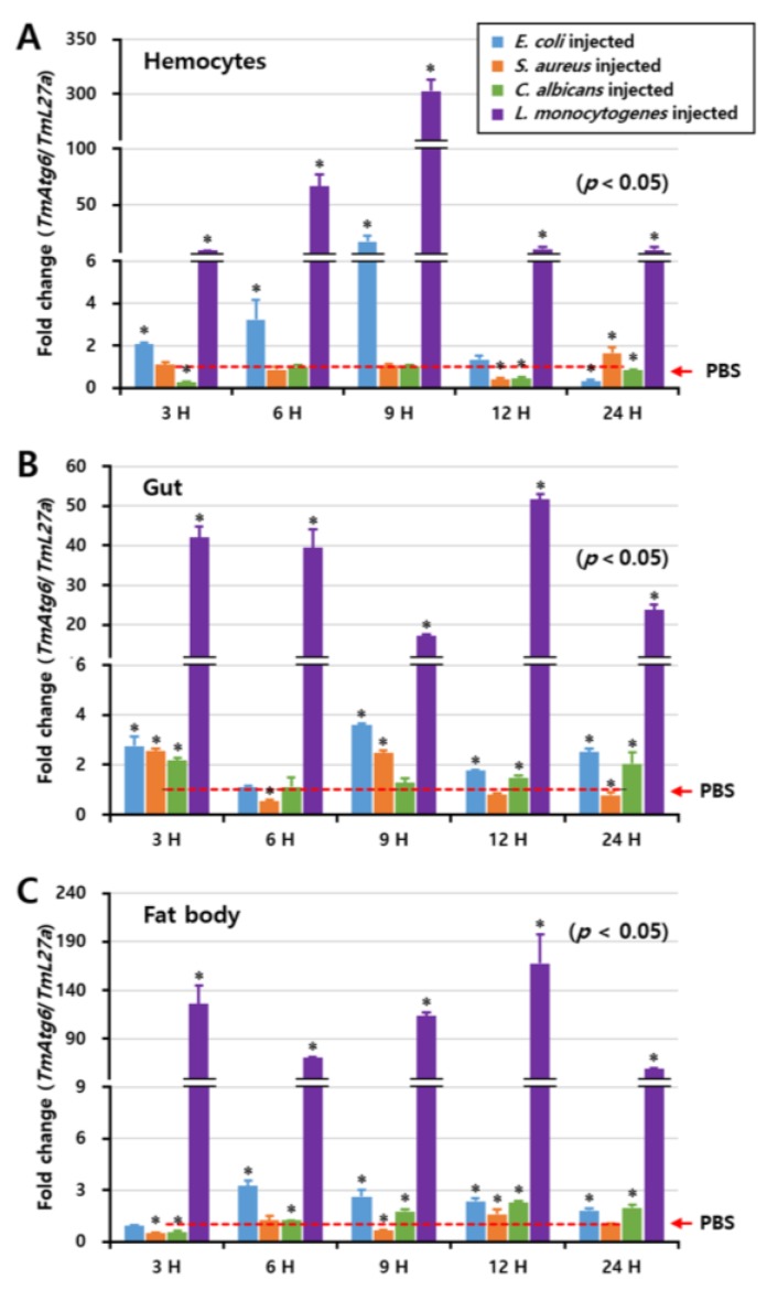
Figure 4.
Induction patterns of TmAtg6 in different tissues against E. coli, S. aureus, C. albicans, and L. monocytogenes, including hemocytes (A), gut (B), and fat body (C). The induction pattern analysis of TmAtg6 gene in different tissues of T. molitor young larvae was performed by injection of E. coli (106 cells/μL), S. aureus (106 cells/μL), C. albicans (5 × 10⁴ cells/μL), or L. monocytogenes (106 cells/μL). Samples were collected at different time points such as 3, 6, 9, 12, and 24 h post-injection of microorganisms. Twenty young larvae of mealworm were used at each time point. In hemocytes, TmAtg6 gene was highly induced at 9 h post-injection of L. monocytogenes. In the gut, the injection of L. monocytogenes highly induced the expression of TmAtg6 at 3 h post-injection, gradually decreased during 6 and 9 h, and then highly induced levels of TmAtg6 12 h post-injection. In the fat body, injection of L. monocytogenes highly induced TmAtg6 at 12 h post-infection.

