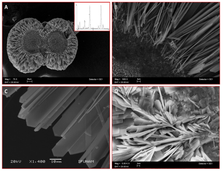
Figure 2.
Scanning electron microscope (SEM) images of sphere-like structures formed in vitro by CEMP1-p4 (inset indicates Ca/P ratio of 1.52) (A). (B) Higher magnification shows an amorphous core with nascent needle-like crystals. (C) Hydroxyapatite crystals show a spear-like form. (D) Control crystals show irradiating crystals and plate-like crystals.

