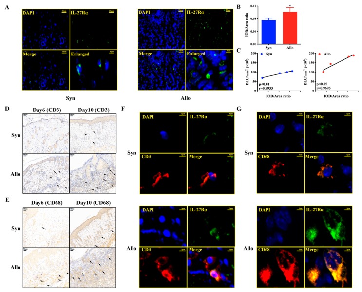
Figure 7.
The relationship of in vivo radioactivity accumulation (125I-anti-IL-27Rα mAb) with IL-27Rα positive cells. Skin transplantation mice models were established and the graft was isolated. (A). The IL-27Rα staining (green) was performed for the graft on day 10 post transplantation, and the cell nucleus was stained with DAPI (blue). (B). The IL-27Rα expression (green) was analyzed and represented by the IOD/Area ratio. (C). Correlation between graft radioactivity uptake (DLU/mm2) and IL-27 Rα expression (IOD/Area ratio) on day 10. (D,E). CD3 (D) and CD68 (E) staining of graft on day 6 and day 10 post transplantation. (F,G). The IL-27Rα (green) expressed on the CD3 (F) and CD68 (G) positive cells of the graft on day 10 post transplantation. * p < 0.05 vs. syngeneic group. Scale bar 20 μm (A), 50 μm (D,E), 5 μm (F,G).

