Figure 4.
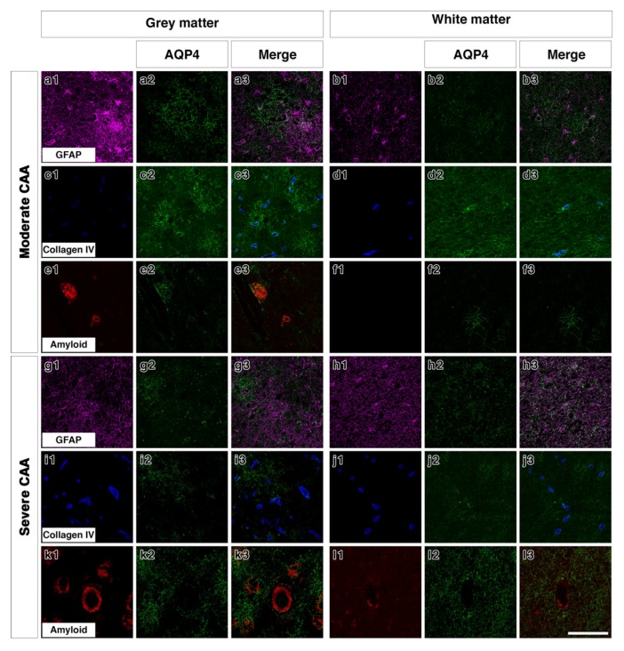
Double immunofluorescence for AQP4 (green) with GFAP (magenta), collagen IV (blue) or Aβ (red) in moderate or severe CAA. In the grey matter of moderate CAA, both AQP4 and GFAP immunostaining appears intense with AQP4 more diffuse (a1–a3). AQP4 appears in a perivascular location as shown by the association with collagen IV (c1–c3) but also appears associated with the presence of Aβ (e1–e3). In the white matter of moderate CAA, AQP4 immunostaining appears fainter (b1–b3), with some close relation to collagen IV (d1–d3). No Aβ immunostaining was detected in the white matter (f1–f3). In the grey matter of severe CAA, immunostaining of GFAP appears more intense than that of AQP4 (g1–g3). Immunostaining of AQP4 (g2) appears reduced when compared to that seen in the grey matter of moderate CAA (a2) but can be observed in a perivascular location, as seen by the relation to collagen IV immunostaining (i1–i3). Immunostaining of AQP4 can also be seen in close proximity to blood vessels laden with Aβ (k1–k3). In the white matter of severe CAA, the immunostaining of AQP4 appears diffuse with some punctate cell bodies stained and little perivascular distribution (h1–h3 and j1–j3). Granular AQP4 immunostaining can be seen surrounding blood vessels containing some Aβ (l1–l3). Scale bar 100 µm.

