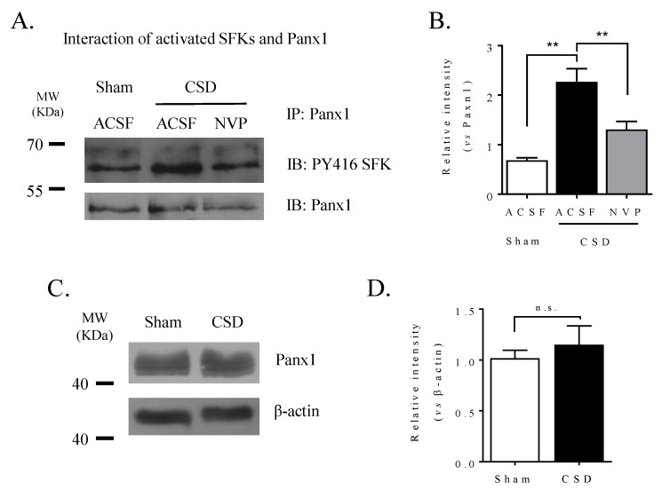
Figure 3.
CSD promoted the interaction of activated SFK with Panx1 channels in the ipsilateral cortex of rat, which was reduced by NVP perfused into the contralateral i.c.v.. (A) Representative images showing co-immunoprecipitation followed by Western blot treated with ACSF or NVP (0.3 nmol) in the absence or presence of 3 M K+-induced CSD. Panx1 was immunoprecipitated (IP) and the pulldown of Panx1 with PY416 SFK was assessed by immunoblotting (IB). Equal loading was indicated by total Panx1 immunoblotting. (B) Quantitative analysis of the IB relative band intensity of PY416 SFK pulled down with Panx1 during IP in cortex treated with ACSF or NVP. n = 5, 6, 4 in the sham, CSD, and NVP+CSD group, respectively. Data were presented as the relative band intensity of PY416 SFK normalized to that of Panx1. (C) Representative images showing immunoblotting of Panx1 with or without CSD induction. (D) Quantitative analysis of the IB relative band intensity of Panx1 treated with ACSF or KCl. n = 3 in the sham and CSD group, respectively. Data were presented as the relative band intensity of Panx1 normalized to that of β-actin. One-tailed, Mann–Whitney test was used for comparisons between sham and CSD group, CSD and NVP group, respectively. All the values shown are means ± SEM. No significant difference was shown as n.s., whereas significant differences were set when * p < 0.05 and ** p < 0.01.

