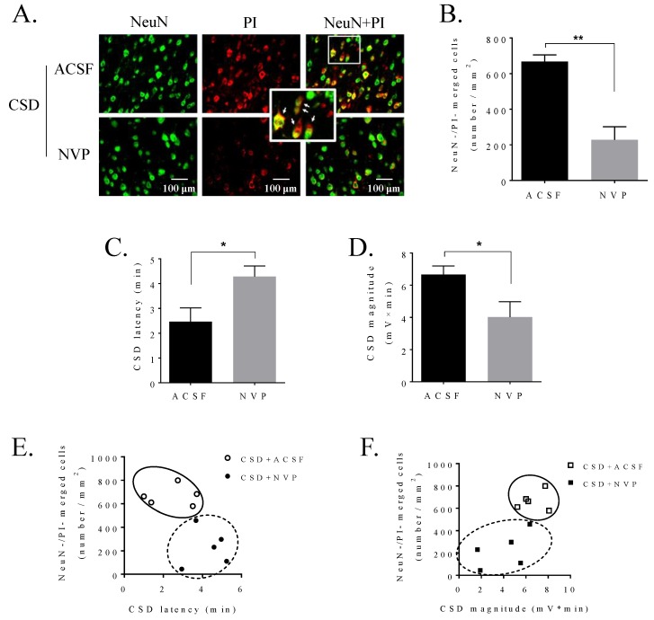
Figure 4.
NVP, the NR2A antagonist, perfused into the contralateral i.c.v. attenuated CSD-induced neuronal PI staining in the ipsilateral cortex of rat and reduced cortical susceptibility to CSD. (A) Representative images of CSD-induced PI staining (red) on NeuN positive cells (green) in layers V and VI of the sensorimotor cortices pretreated with ACSF or NVP. PI and NeuN positive cells (yellow) were pointed by arrows in the inset. (B) Neuronal PI staining was indicated by the number of merged cells (cells/mm2) after pretreatment of ACSF (n = 5) or 0.3 nmol NVP (n = 5) and CSD induction in sensorimotor cortices of rats. (C,D) Effects of 0.3 nmol NVP on CSD latency (minute) and magnitude (mV × minute) (n = 5 for each group). (E,F) Correlation between the level of PI staining and CSD latency/CSD magnitude pretreated with ACSF or 0.3 nmol NVP (n = 5 for each). One-tailed, Mann–Whitney test was used for comparison between ACSF and NVP groups. All values are shown as means ± SEM. Significant differences were set when * p < 0.05 and ** p < 0.01.

