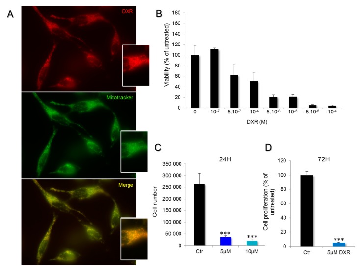
Figure 1.
Incorporation of doxorubicin (DXR) in mitochondria of HeLa cells and impact on cell viability. (A) Living cells were treated with 100 nM Mitotracker Green and 5 μM DXR, for 30 and 10 min, respectively. The overlaps between mitochondria (green) and DXR (red) are shown in yellow. (B) The viability of HeLa cells was estimated after doxorubicin treatment for 24 h with a range of concentrations between 10−7 and 10−4 M. (C) The proliferation was measured, after 24 h with DXR (5, dark blue and 10 μM, light blue), by counting the cells in the presence of trypan blue, and it was compared to the condition without DXR, the control (Ctr, black). ***, p < 0.001; One-way ANOVA with multiple comparisons. (D) The effect of DXR on cell proliferation was also measured after 72 h of treatment and compared to the control untreated cells. ***, p < 0.001; unpaired t test, two-tailed.

