Figure 2.
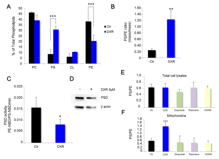
DXR modifies mitochondrial membrane composition of HeLa cells while inhibiting phosphatidylserine decarboxylase (PSD) activity. The effect of DXR on the phospholipid composition of mitochondrial membranes was analyzed by high-performance thin-layer chromatography. (A) The quantification of PC, phosphatidylcholine; PS, phosphatidylserine; CL, cardiolipin; and PE, phosphatidylethanolamine was performed by densitometry and expressed as a percentage of total phospholipids. The mitochondrial membrane composition obtained after 5 μM DXR treatment was compared to that in control conditions. ***, p < 0.001; Multiple t test of two-way ANOVA. (B) The PS/PE ratio was determined in HeLa cells, treated or untreated, with DXR at 5 μM (**, p < 0.01). (C) The rate of PSD activity was determined in vitro on isolated mitochondria using a fluorescent analog of phosphatidylserine, PS-NBD (black). The enzyme activity was also measured in the presence of DXR 5 μM (blue). The data are expressed in PE-NBD/PS-NBD ratio per minute. Each point represents the mean value ± SEM of N ≥ 3 experiments (*, p < 0.05). (D) The expression level of PSD was analyzed by Western blot on cell lysate treated by 5 μM DXR for 24 h, and compared to the untreated cells. The effect of topoisomerase inhibitors on PS/PE ratios was determined from (E) total cell lysates and the (F) isolated mitochondrial fraction. Cells were incubated with 5 μM DXR, 1.25 μM etoposide, 20 μM razoxane, or 10 μM XK469 for 24 h. Then, estimation of the PS/PE ratios was performed. The data are expressed as mean values ± SD with ***, p < 0.001.

