Figure 3.
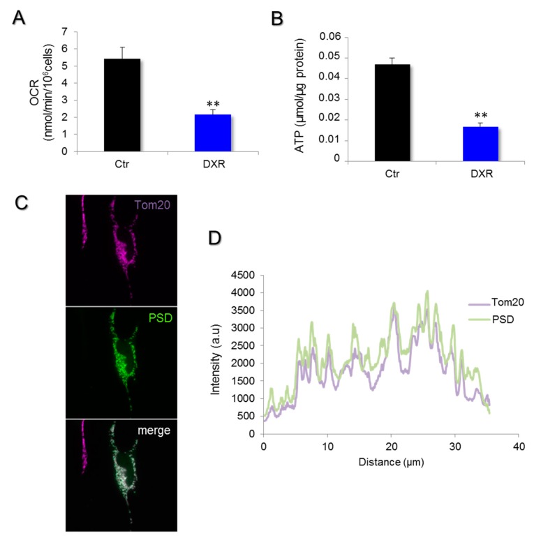
Doxorubicin inhibits mitochondrial respiration and reduces ATP content. (A) Mitochondrial respiration was measured in HeLa cells treated for 6 h with 5 μM DXR, and the endogenous rate of respiration was recorded. Data are expressed as mean values of oxygen consumption rates (OCRs) ± SEM, N = 5 experiments with p = 0.006 (**, p < 0.01). (B) The total ATP content of HeLa was determined by bioluminescence in cells treated or untreated with 5 μM doxorubicin for 24 h. The results are expressed in μmol/μg protein as mean values ± SEM. The difference was considered as significant when p < 0.05 (**, p < 0.01). (C) The PSD subcellular repartition was investigated by immunofluorescence on HeLa cells. Cells were transfected for 48 h with pCMV6-PSD-DDK-myc plasmids and were then fixed with PFA 4%. After cell permeabilization by triton 15%, PSD was labeled with the anti-DDK antibody and the secondary antibody AF-488 anti-mouse (green). Immunostaining of the mitochondrial network (far red) was obtained with an anti-TOM20 antibody and a secondary one, AF-647 anti-rabbit. (D) The fluorescence intensity was measured and illustrated with the line-scan to show the overlaps between Tom20 (far red) and PSD expression (green).

