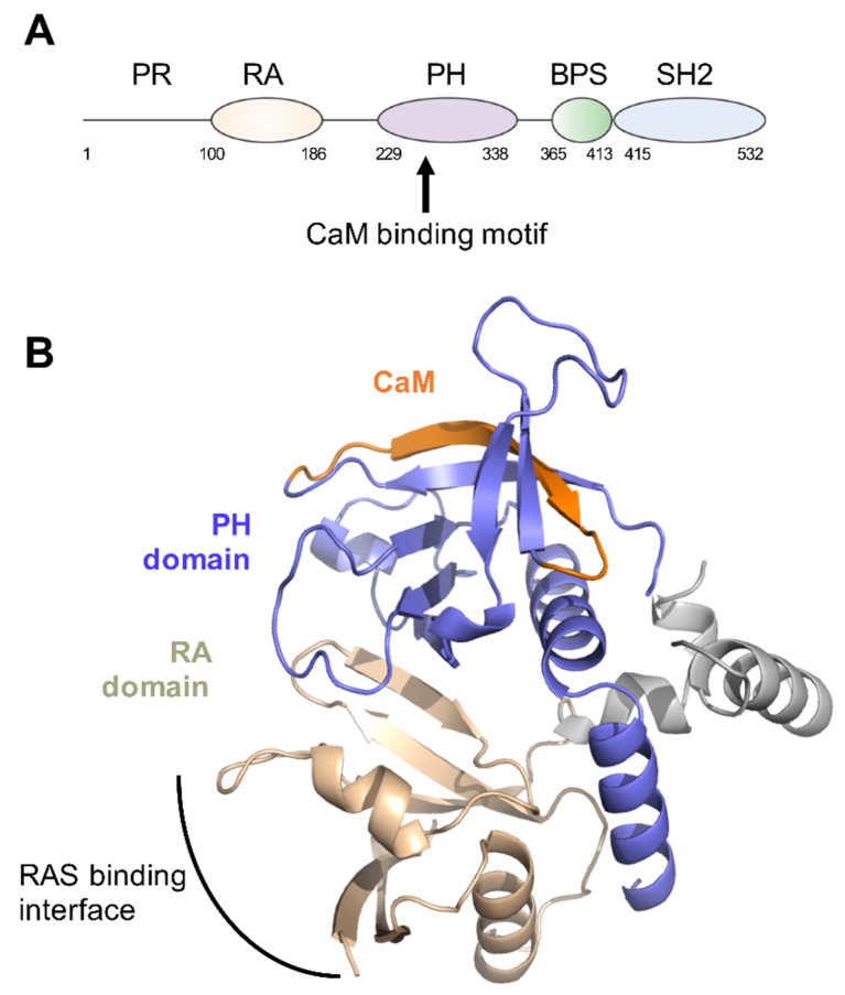
Figure 1.
Grb7 domain structure. (A) Schematic depicting the arrangement of Grb7 domains and highlighting the position of the postulated calmodulin (CaM) binding site; (B) Model of the Grb7 RA-PH domains based upon the Grb10 RA-PH structure (PDB:3HK0). The RA domain is coloured wheat, the PH domain is purple and the residues that correspond to the Grb7 CaM-BD are coloured orange.

