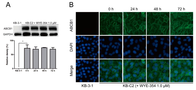
Figure 6.
Expression level and subcellular localization of ABCB1 after up to 72 h of treatment with WYE-354. (A) Western blotting on the expression level of ABCB1 in the KB-C2 cells incubated with 1.0 µM WYE-354. Image J was used to quantify the relative density of each band. * p < 0.05, compared with the control group. (B) Immunofluorescence on the subcellular localization of ABCB1 in the KB-C2 cells treated with 1.0 µM WYE-354. Green—ABCB1. Blue—4′,6-diamidino-2-phenylindole (DAPI) counterstains the nuclei.

