Figure 2.
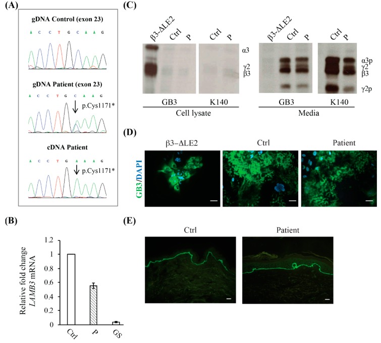
Molecular characteristics of the patient. (A) LAMB3 exon 23 sequence chromatogram of patient genomic DNA (gDNA) shows the p.Cys1171* compound heterozygous mutation (middle panel). A sequence chromatogram from a normal control is shown for comparison (upper panel). The sequence of a complementary DNA (cDNA) fragment reverse-transcribed from the total RNA of patient keratinocytes reveals the p.Cys1171* mutation as unique peak, indicating that the mRNA carrying this variant is stable and contributes alone to protein synthesis (lower panel). (B) Real-time RT-PCR shows that LAMB3 total RNA levels in patient keratinocytes (P) are reduced to almost 50% of a normal control (Ctrl), while they are dramatically decreased in a junctional epidermolysis bullosa generalized severe keratinocyte strain (GS) carrying premature termination codon mutations in both LAMB3 alleles. (C) Immunoprecipitation of laminin-332 (LM332) from cell lysates and media of 35S-labelled keratinocytes from our patient (P), a previously reported junctional epidermolysis bullosa generalized intermediate (JEB-GI) individual (β3-ΔLE2) with a mutation causing an unfolded β3 short arm submodule, and a normal control (Ctrl). Equal amounts of protein-bound radioactivity were immunoprecipitated with mAbs GB3 and K140 and loaded on the gels under reducing conditions. Increased levels of unprocessed LM332 are only detected in cell lysates from the β3-ΔLE2 individual (using GB3), while, in comparison, samples from our patient and normal control show barely detectable levels of the unprocessed protein (using GB3 and K140) (left panel). Conversely, the mature LM332 is barely detected in β3-ΔLE2 spent medium and highly abundant in the media from the normal control and patient keratinocytes. The levels of processed LM332 were approximately 60% in comparison to normal cells. (D) Immunofluorescence localization of LM332 in junctional epidermolysis bullosa (JEB) keratinocytes. The GB3 antibody (against heterotrimeric LM332) stains the protein extracellularly in the patient and control keratinocytes (middle and right panel). In contrast, staining was almost exclusively confined within the cell cytoplasm in the β3-ΔLE2 cells (left panel). Scale bars = 10 µm. (E) Immunofluorescence of patient skin (right panel) using GB3 antibody shows a positive staining almost comparable to control skin (left panels). Scale bars = 20 µm.

