Figure 3.
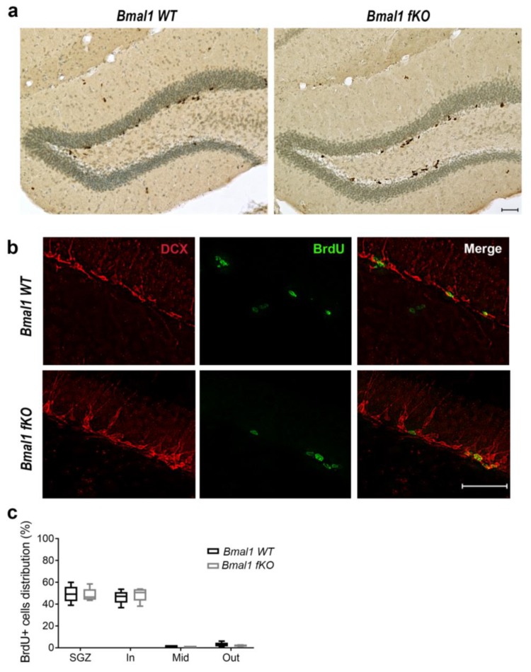
The proliferation and spatial distribution of neural progenitor cells in the dentate gyrus of Bmal1 fKO mice. (a) Representative photomicrograph of Bromodeoxyuridine-positive (BrdU+) cells (brown) and cresyl violet staining (blue) in the dentate gyrus of Bmal1 WT and Bmal1 fKO mice. Scale bar = 50 µm. (b) Representative immunofluorescence of the marker for proliferating cells BrdU+ (green) and the marker for neuronal precursors/migrating neuroblasts, doublecortin (DCX) (red). (c) Quantification of BrdU+ cells within the subgranular zone (SGZ), the inner (In), middle (Mid) or an outer (Out) third of the granular cell layer of the dentate gyrus. n = 5 of each genotype.

