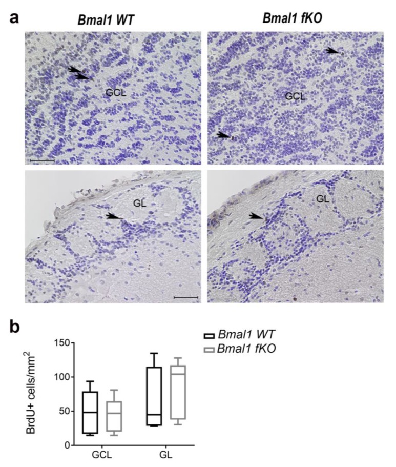
Figure 7.
Proliferating cells in the olfactory bulb of Bmal1 fKO mice. (a) Representative photomicrographs of BrdU+ cells (brown, arrows) and cresyl violet staining (blue) in the granule cell layer (GCL) and the glomerular layer (GL) of the olfactory bulb. Scale bars = 50 µm. (b) The number of BrdU+ cells in the GL and the GL of the olfactory bulb was not different between Bmal1 WT and Bmal1 fKO mice. n = 5 mice per genotype.

