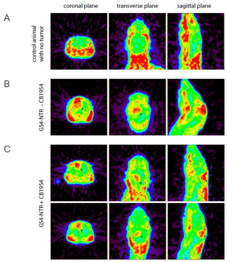
Figure 5.
MicroPET imaging of intracerebral RG-2 glioma-bearing rats. MicroPET imaging of L-[18F] FET in rats was performed using the R4 system, 20 days after intracerebral RG-2 glioma implantation. Representative examples comparing microPET imaging results from non-tumor-bearing rats (A), tumor-bearing rats treated with GS4-NTR but without prodrug administration (B), or tumor-bearing rats with GS4-NTR and CB1954 prodrug treatments (C) are shown, and tumor regions are indicated by arrowheads. The tumor uptake of L-[18F] FET in the GS4-NTR−CB1954 group relative to the GS4-NTR + CB1954 group was 2.015-fold (p < 0.01).

