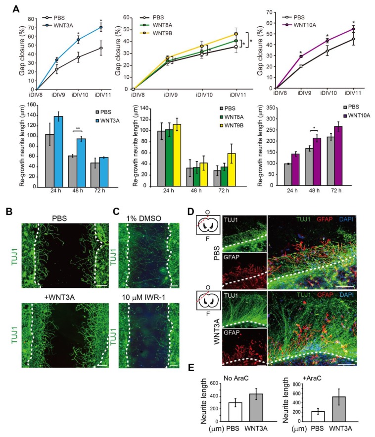
Figure 3.
Recombinant WNT3A promotes regeneration of injured cortical neurons and brain tissues. (A) Cortical neurons were pre-treated with PBS, WNT3A, WNT8A, WNT9B, or WNT10A recombinant proteins prior to injury on DIV8. Upper: The percentages of gap closure were calculated from iDIV8 to iDIV11. Bottom: The lengths of re-growing neurites were measured between 0–24 h, 24–48 h, and 48–72 h after injury. Data are presented as mean ± SEM (n = 4 for WNT3A and WNT9B experiments; n = 5 for WNT10A experiment). * p ≤ 0.05, ** p ≤ 0.01 (paired Student’s t-test). (B) Cortical neurons with or without WNT3A (50 ng/mL) pre-treatment were fixed and subjected to immunofluorescence staining with anti-TUJ1 antibody (neurites) on iDIV11. (C) Cortical neurons were treated with 1% DMSO or IWR-1 prior to injury on DIV8. Neurons were subjected to immunofluorescence staining with anti-TUJ1 antibody on DIV11 and the representative images are shown. Dashed lines in (B,C) indicate the borders of the injured gap. Scale bar, 100 µm. (D) The effect of WNT3A on neuronal regeneration was assessed using organotypic brain slice culture. Brain slices were injured at the olfactory tubercle on DIV0, as indicated by the red dashed line in the upper left atlas map, and cultured with or without WNT3A (50 ng/mL) in AraC-containing medium. Brain tissues were fixed on iDIV4 and subjected to immunofluorescence staining with anti-TUJ1 (green) and anti-GFAP (red, glia) antibodies, and DAPI (blue). The border of injured sites is depicted by white dashed lines, and remaining brain tissue is underneath the lines in each panel. F: Frontal lobe; O: Occipital lobe. Scale bar, 100 µm. (E) The length of regenerating neurites from the brain slices treated with PBS, WNT3A, AraC + PBS, or AraC + WNT3A were calculated. Data are presented as mean ± SEM (n = 4). Data were analyzed using non-parametric Mann–Whitney test.

