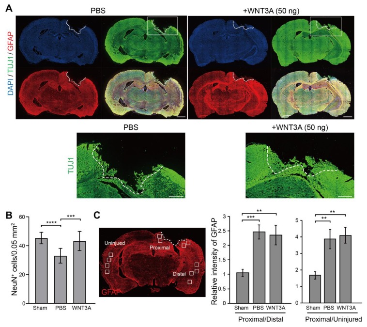
Figure 4.
Recombinant WNT3A promotes regeneration of injured brain tissue of controlled cortical impact (CCI) mice. (A) The effect of WNT3A on neuronal regeneration was assessed using in vivo CCI mouse model. Cortical impact on motor and somatosensory cortex was generated and the mice were given PBS or 50 ng WNT3A intranasally during 0–3 dpi. Injured brain tissues were fixed, cryosectioned on 4 dpi, and subjected to immunofluorescence staining with anti-TUJ1 (green), anti-GFAP (red) antibodies, and DAPI (blue). White dashed lines depict the border of CCI sites. The regions of interest (ROIs) are indicated in the boxed regions. Scale bar, 1 mm. Enlarged images of the ROIs are shown at the bottom. Scale bar, 0.5 mm. (B) The number of NeuN+ cells and (C) the relative intensity of GFAP of CCI cryosections from sham group (n = 10), PBS-treated (n = 12) and WNT3A-treated (n = 8) CCI animals were quantified as described in the Materials and Methods. GFAP-immunostained cryosection on the left panel in (C) defines 0.05 mm2-ROIs within white boxes. Dashed lines indicate the borders of the impact site. Data are presented as mean ± SEM. ** p ≤ 0.01, *** p ≤ 0.001, **** p ≤ 0.0001, analyzed by one-way ANOVA followed by Tukey’s test.

