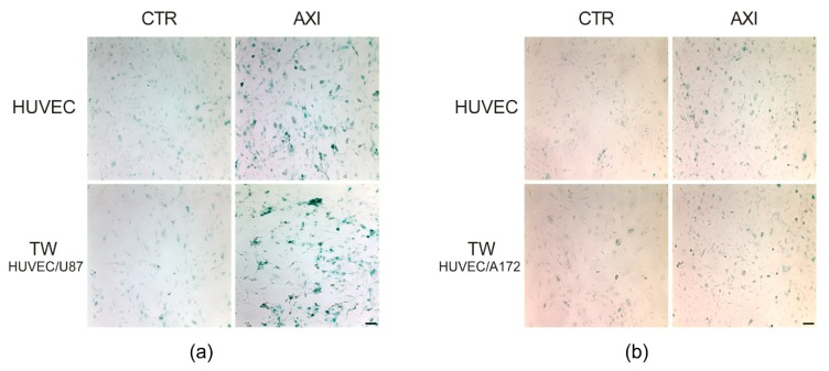
Figure 2.
Axitinib-dependent SA-β- galactosidase staining of HUVECs wasnot altered by GBM cells coculture. Four days after Axitinib pulse, HUVECs appeared positive to β-galactosidase staining, with a typical blue color. This effect was not altered in HUVECs cocultured for 48 h in transwell with GBM cell lines, U87MG (a) or A172 (b). n = three biological replicates. Magnification 10×, scale bar 100 µm.

