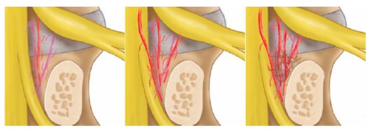
Figure 8.
Drawings of the neovascularization grading system. Left picture showing a grade 1 normal appearance with sparse epidural vessels around the disc. The middle picture showed a grade 2 increased neovascularization of epidural vessels with vascularization. The right picture showed grade 3 with increased neovascularization of epidural vessels with vascularization and adhesion on neural tissues.

