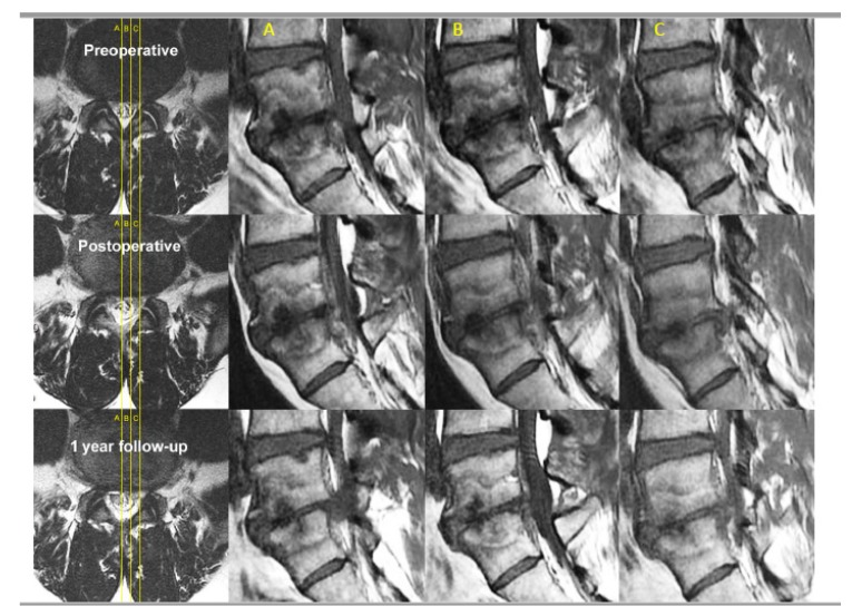
Figure 9.
Sagittal cuts across L5/S1 in a patient with spinal canal stenosis, degenerative disc disease of L5/S1 with disc protrusion into the spinal canal and vertebral bodies of L4 and L5, there are Type 1 Modic changes signifying bone marrow edema and inflammation. The corresponding axial and sagittal cuts are labelled (A–C) accordingly. On axial cuts, we can see preoperative spinal canal stenosis with disc bulge and ligamentum hypertrophy with Schiaz grade A3 with rootlets lying dorsally occupying more than half of the dura sac area. This patient suffered buttock pain and both leg pain especially right leg pain for 5 months. The patient visited the clinic with visual analogue scale (VAS) score of 8 that was aggravated from 3 months ago, especially buttock pain and right leg pain, in spite of the preoperative MRI and spinal stenosis was not severe (Schiaz Grade A3). We can check the severe Modic change (Type 1) in the adjacent vertebrae. The patient had decompressed and radiofrequency ablation to sinuvertebral and basivertebral nerves in MRI postoperative day 1 which had been maintained in a 1-year follow up. There is shrinkage of the disc and less marrow changes in bone edema. The patient’s final follow-up VAS was 1 and returned to work as per normal.

