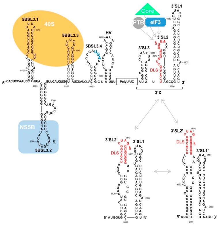
Figure 5.
The 3′ end of the HCV genome. This figure shows the sequence and the widely accepted secondary structure model of the genomic 3′UTR and the upstream functional region containing the 5BSL3.1, 5BSL3.2, 5BSL3.3 and 5BSL3.4 domains. The theoretical alternative conformations acquired by the 3′X tail are also shown. The palindromic motif involved in HCV genome dimerization (DLS, dimer linkage sequence) is shown in red. The k and k’ sequences in the DLS and apical loop of the 5BSL3.2 domain respectively are underlined; these are required by both domains for their interaction activity. The translation stop codon is shown by enlarged blue lettering. The binding sites for viral and cellular proteins are indicated by colored backgrounds. Nucleotide numbering is as in Figure 3.

