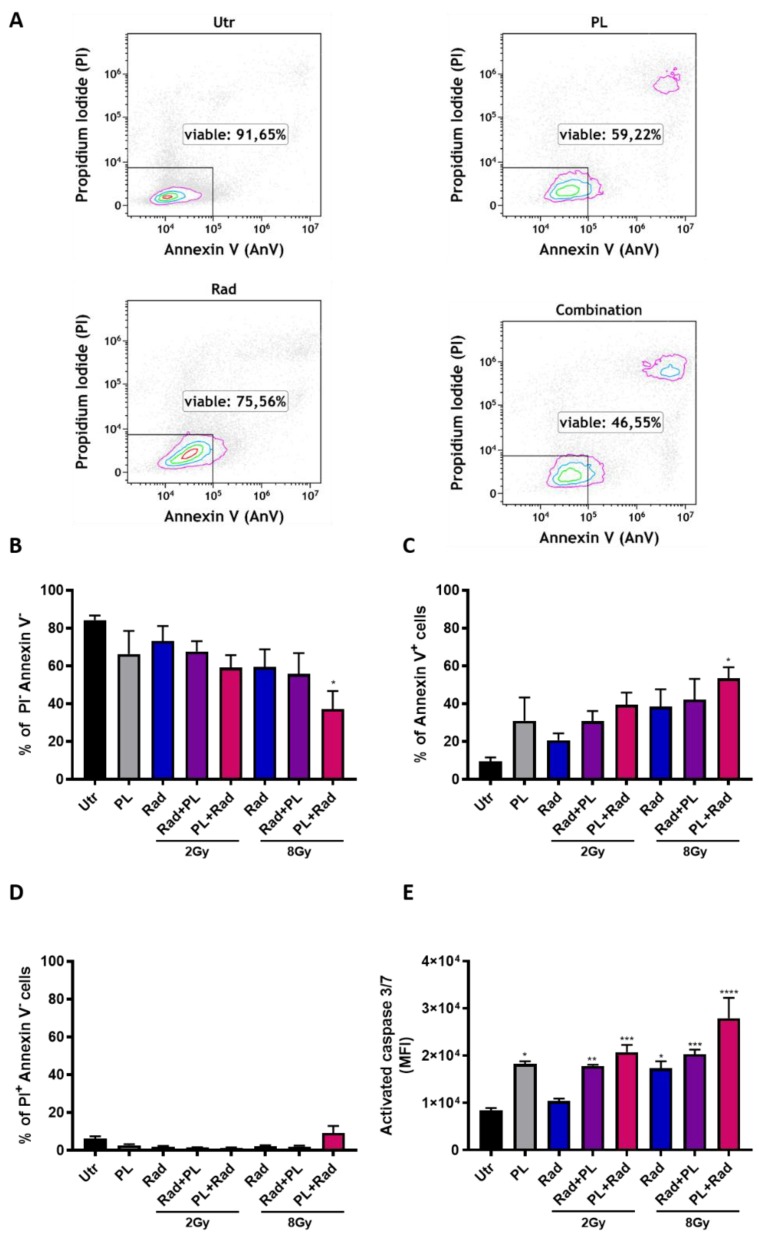
Figure 2.
Gas plasma and single-dose radiotherapy induced apoptosis in an additive fashion. (A) representative dot plots of Annexin V and propidium iodide (PI) flow cytometric analysis in B16F10 cells 24 h post-exposure to plasma (15 s), radiotherapy, or both; (B–D) quantification of viable (B), apoptotic (C), and necrotic (D) cells; (E) additional confirmation of apoptosis using the cell event caspase 3/7 dye. Data are representative and mean + SE of three experiments. Statistical analysis was performed by one-way ANOVA. Gy = gray, Utr = untreated, PL = plasma, Rad = radiotherapy, MFI = mean fluorescent intensity; * = p < 0.05, ** = p < 0.01, *** = p < 0.001, **** = p < 0.0001.

