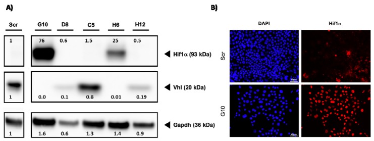
Figure 1.
Loss of von Hippel–Lindau (VHL) protein induces nuclear Hif1A expression. (A) Cell lysates from control cells (Scr) and 5 VHL clones were prepared and the expression of VHL and Hif1a was analyzed by Western blot. An antibody directed against Gapdh served as control. The numbers indicate ratios in signal intensity compared to Scr. (B) Cells were cultivated on glass coverslips. After fixation, the cells were incubated with a specific Hif1a antibody. A secondary Alexa-568 labeled antibody was used to visualize the signals. The staining of the nuclei was done by incubation with 4′,6-diamidino-2-phenylindole (DAPI) (scale bar = 40 µm).

