Figure 2.
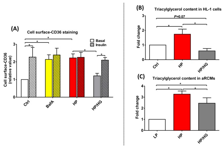
Cell surface-CD36 staining and triacylglycerol contents in lipid-overexposed cardiomyocytes. (A) Cell surface CD36 staining of HL-1 cells: Prior to CD36 cell surface staining, HL-1 cells were treated for 24 h either with control (Ctrl) medium, Ctrl medium containing 100 nM Bafilomycin-A (BafA), high palmitate medium containing 500 µM palmitate and 100 nM insulin (HP), or HP medium with 25 mM glucose (HP/HG). Subsequently, cells were stimulated either without or with 200 nM insulin for 30 min and immunochemically stained for cell surface CD36 content (n = 3). (B-C) Triacylglycerol contents in lipid-overexposed cardiomyocytes: (B) HL-1 cells were incubated for 24 h with either Ctrl medium, HP medium, or HP/HG (n = 5); (C) aRCMs were incubated for 24 h with either LP medium, HP medium, or HP/ HG (n = 7). Values are displayed as mean ± SEM. * p < 0.05 were considered statistically significant.

