Figure 5.
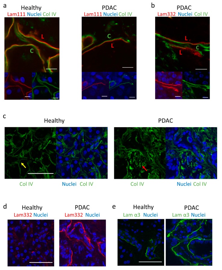
Composition of basement membrane in healthy and PDAC patients. (a) Immunofluorescence images of laminin/collagen IV bilayer in healthy and PDAC patients. Scale bar = 10 µm. L = laminin 111-rich side. C = collagen IV-rich side. Upper panel = Lam111 + Col IV, lower left = Lam111 + DAPI, lower right = Col IV + DAPI. (b) Immunofluorescence images of laminin 332/collagen IV bilayer in PDAC. Scale bar = 10 µm. L = laminin 111-rich side. C = collagen IV-rich side. Upper panel = Lam332 + Col IV, lower left = Lam332 + DAPI, lower right = Col IV + DAPI. (c) Immunofluorescence images of collagen IV organisation in healthy and PDAC tissues. Scale bar = 50 µm. Yellow arrow indicates organised collagen IV, red arrow indicates disorganised collagen IV. (d) Immunofluorescence images of laminin 332 organisation in healthy and PDAC tissues. Scale bar = 50 µm. (e) Immunofluorescence images of laminin α3 organisation in healthy and PDAC tissues, using P3H9 monoclonal antibody. Scale bar = 50 µm.

