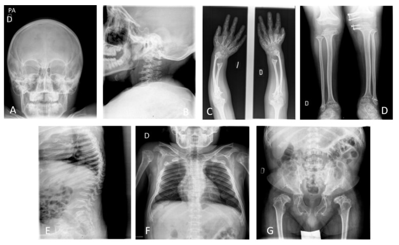
Figure 2.
X-ray images of a 17-year-old male patient with MPS IVA. Images show skeletal dysplasia (dysostosis multiplex); (A) incomplete ossification and prominent forehead, (B) incomplete ossification in odontoid process and subluxation of the atlas secondary to odontoid hypoplasia with platyspondyly of cervical vertebrae, (C) cortical thinning and mild widening of the diaphysis of the humerus and tilting of the radial epiphysis towards the ulna producing curvature, (D) genu valgum with cortical thinning of tibia and fibula, (E) the accentuated dorsal thoracolumbar kypholordosis with the advanced platyspondyly, irregularity, and anterior beaking of vertebral bodies characteristic of MPS IVA and flared ribs, (F) abnormal thoracic cage, pectus carinatum and scoliosis with oar shaped ribs: the ribs are wide anteriorly and laterally and overconstricted in their paravertebral portions, (G) spondyloepiphyseal dysplastic femoral heads and oblique acetabular roof with coxa valgus deformity and flared iliac wings.

