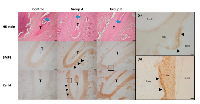
Figure 6.
Histological analysis under the hematoxylin and eosin (HE) stain. The direction of tooth movement is indicated by the blue arrow at the central root of the first molar (T). The width of the periodontal ligament space was narrower on the compression side than on the tension side. The expression level of bone morphogenic protein-2 was higher in Group A and B compared to the control group. The expression level was higher on the tension side (arrow heads). The expression level of RANKL was also higher in Group A and B compared to the control group. The expression level was higher on the compression side (arrow heads). (a) High magnification views for BMP2 of Group B showed the expression was mainly found in the cells lined on the tension side of the alveolar bone (arrow heads, original magnification ×400) (PDL: periodontal ligament). (b) High magnification views for RANKL of Group A showed the expression was mainly found in the cells lined on the compression side of the alveolar bone (arrow heads, original magnification ×400).

