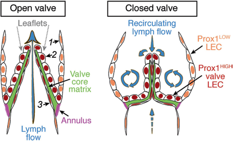
Fig. 1.
Lymphatic valve organization and the associated lymph flow patterns. The leaflet is covered on both sides by Prox1HIGH LECs, which are more compact on the sinusal side subjected to recirculating flow and more elongated on the luminal side subjected to high laminar shear stress [1, 2]. A core of specialized ECM is sandwiched between the two layers of LECs. Leaflets are inserted within the vessel wall at the annulus, a fibrous circumferential fold rich in collagens and elastin, which changes shape during the valve cycle [3].

