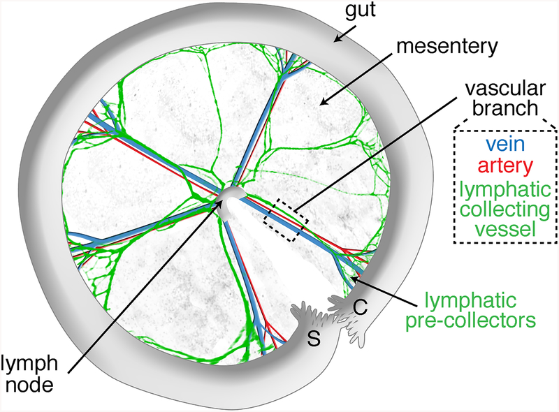
Fig. 2.
Scheme of a dissected anterior mesentery, unfolded clockwise from duodenum (Duo) to ileum highlighting its main components: intestine, mesentery, lymph node, and five vascular branches. The black dashed box indicates the vascular branch area selected for lymphatic vessel length and valve quantifications. The orange and pink dashed lines indicate the vascular branch segment that is selected for analysis of valves from collecting vessels and precollectors, respectively.

