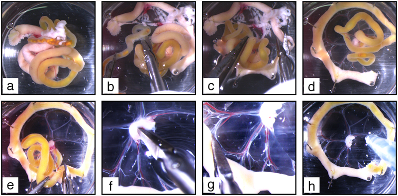
Fig. 6.
Mesentery dissection procedure. (a) The duodenum is pinned on elastomere to allow further clockwise unfolding of the intestine. (b) Clockwise unfolding gives access to the colon, which is excised above the caecum. (c) The pancreas (white diffuse tissue spread in the mesentery at the top of the image) is excised. (d) The mesentery is further unfolded to make a complete wheel. (e) The remaining part of the intestine is cut off. (f) The upper half of the lymph node is excised. (g) Small holes are made at the connection of each vascular branch with the intestine. (h) Blood and chyle are flushed out from mesenteric vessels

