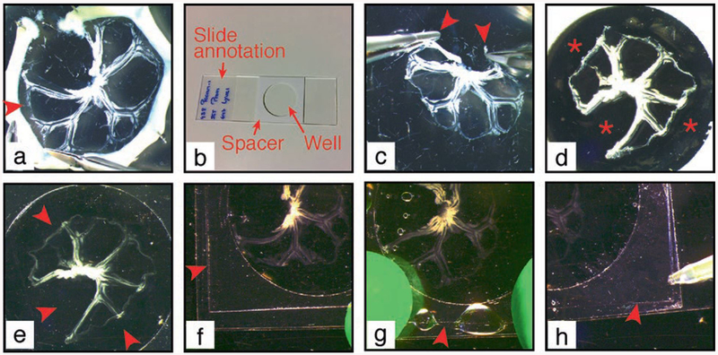
Fig. 8.
Procedure of mesentery mounting on microscopy slide. (a) The intestine is cut off the mesentery at the junction between the two. Arrowhead: detachment of the mesentery (b) Microscopy slide is prepared with a central spacer. Mesentery is grasped at the two external extremities (arrowheads) (c) and transferred to the slide within the spacer well, when the opaque liner is still present (d). Asterisk: empty well. (e) Opaque liner is removed and mesentery is covered with mounting medium to fill in the spacer well. Arrowheads: well filled with mounting medium. A coverslip (arrowhead) is applied to a sticky border of the spacer (f), let to fall on the mesentery and tightly attached to the spacer (g). (h) Mounting medium is added to remove air bubbles under the coverslip

