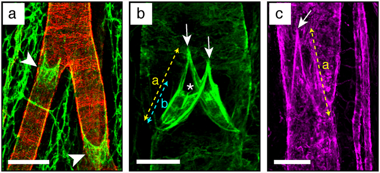
Fig. 9.
Examples of mesenteric lymphatic vessels and valve ECM stainings. (a) Collecting vessel bifurcation stained to analyze its coverage by mural cells. Pecam1 in green, alphaSMA in red. Scale bar: 100 μm. (b) Valve leaflet core matrix stained for laminin alpha5. Scale bar: 30 μm. (c) Valve matrix stained for collagen IV. Scale bar: 50 μm. Arrowhead, lymphatic valve devoid of mural cells; arrow: valve buttress; asterisk: luminal free edge of the valve leaflets; (a) valve length; (b) leaflet length

