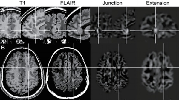
Fig. 4.

Application of a voxel-based image postprocessing method. (A) Right frontomesial focal cortical dysplasia (FCD) was not detected by conventional visual analysis; however, it was clearly visible on the junction image (blurred gray-white matter junction) and extension image (gray matter extending abnormally into the white matter). (B) FCD IIa located at the bottom of the left superior frontal sulcus with a moderate abnormality visible on the junction image; however, no abnormality was visible on the extension, T1-weighted, or fluid-attenuated inversion recovery images. Adapted from Wagner et al. Brain 2011;134(Pt 10):2844-54, with permission from Oxford University Press [45].
