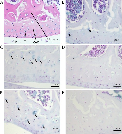
Figure 2.

Initiation and progression of ochronosis in Hgd tm1a mice. H&E staining in A shows division of articular cartilage into different zones: hyaline cartilage (HC) and calcified articular cartilage (CAC) separated by the tidemark (T). Deep to calcified articular cartilage is subchondral bone (SB). B–F show femoral condyles from Hgd tm1a mice that have been Schmorl’s-stained (stains ochronotic pigment a blue colour). B shows pericellular pigmentation of chondrons situated in the calcified articular cartilage in a 9-week Hgd tm1a −/− femoral condyle. C and E show the femoral condyle of Hgd tm1a −/− mice at 26 and 40 weeks, respectively. The pigmentation has advanced to the inner compartment of the cell, with more numerous affected chondrocytes showing varying pigment intensities. The pigmentation is still confined to the calcified articular cartilage layer. Hgd tm1a −/+ mice at 26 and 40 weeks (D and F respectively) show no pigmentation. All sections: 6 μm.
