Figure 1.

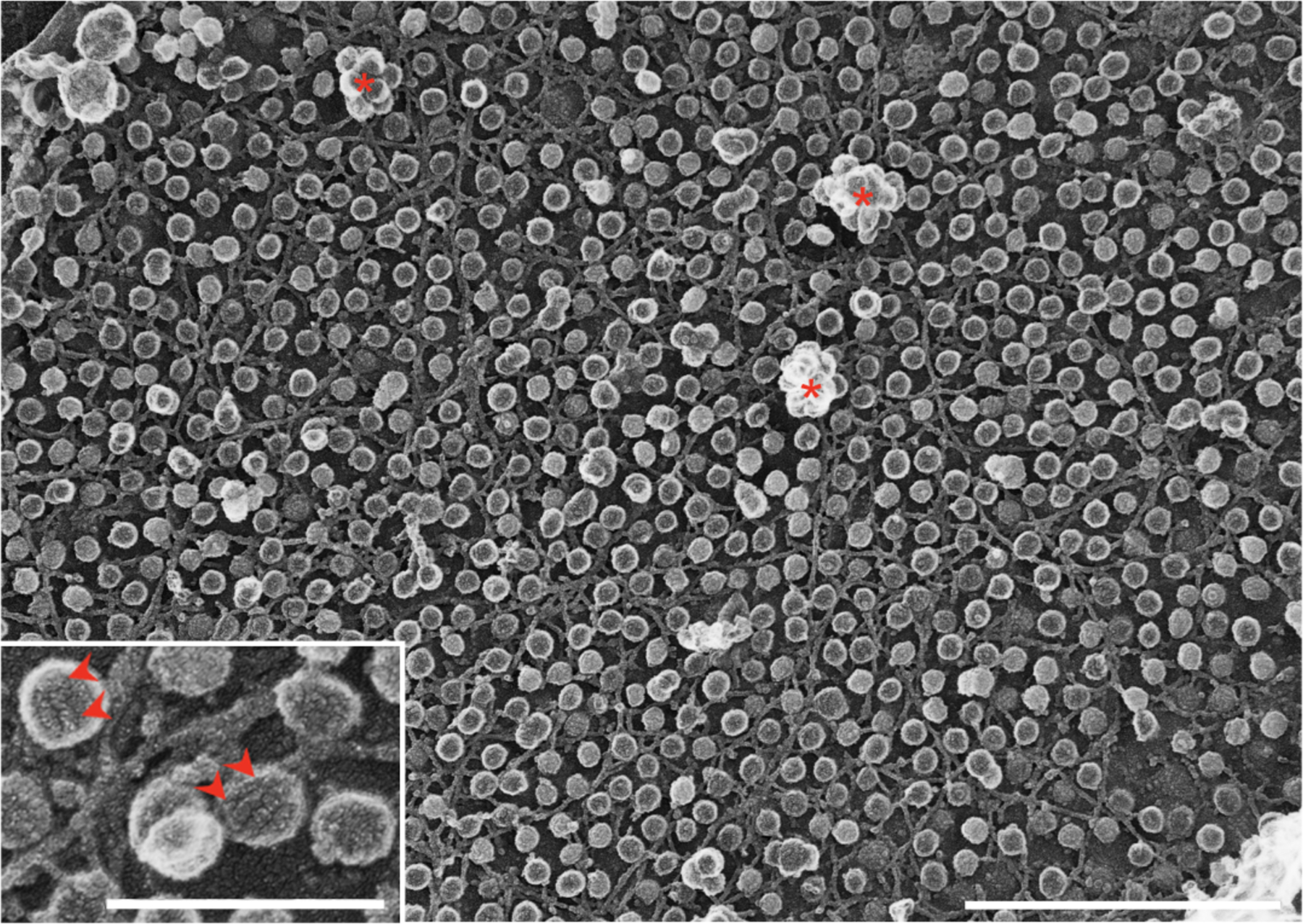
Platinum-replica EM of the plasma membrane of cultured myotubes showing abundant caveolae; both single caveolae and caveolar clusters or rosettes (asterisks) are evident. The characteristic striped coats of caveolae can be observed at higher magnification (inset; arrowheads). Bars, main figure 1μm; inset 0.25μm.
