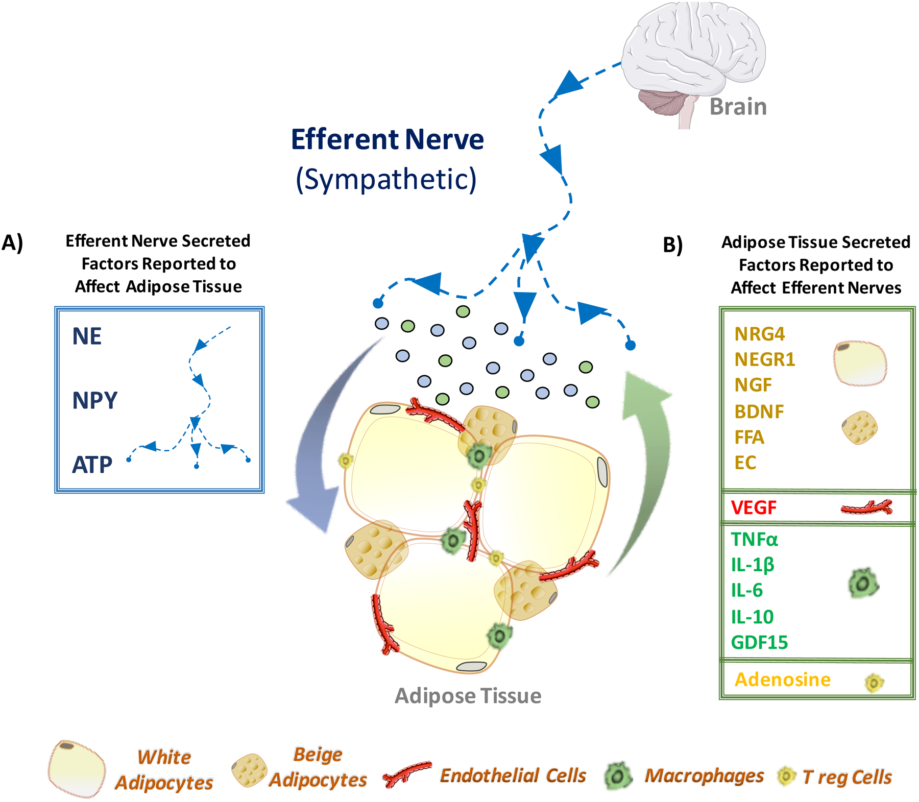
Figure 5: Adipose Tissue-Sympathetic Nerve Crosstalk.

(A) Efferent nerve fibers are known to regulate adipose tissue functions through secretion of bioactive factors. Among them are catecholamine (norepinephrine), neuropeptide Y (NPY) and adenosine triphosphate (ATP). The central nervous system stimulates sympathetic outflow to adipose tissue, triggering the secretion of norepinephrine, NPY or ATP (represented by blue dots). These sympathetic-derived secreted factors, through the activation of their respective receptors, affect not only adipocytes, but also other adipose resident cells, such as endothelial cells, macrophages and lymphocytes. (B) In turn, the stimulated adipose cells also produce a number of secreted bioactive factors that communicate with adipose sympathetic fibers. For instance, multilocular beige adipocytes - induced via sympathetic norepinephrine and/or adenosine molecules - produce the neurotrophic factor neuregulin-4 (NRG4), which is known to promote neurite outgrowth. Additionally, white adipocytes have been shown to synthesize several factors with neurotrophic activity that may enhance sympathetic innervation of adipose tissue. Among these factors are the neuronal growth regulator 1 (NEGR1), nerve growth factor (NGF), brain-derived neurotrophic factor (BDNF), free fatty acids (FFA) and endocannabinoids (EC). The vascular endothelial growth factor (VEGF) secreted by endothelial cells and adipocytes elicits sympathetic innervation in adipose tissue. Adipose resident immune cells, such as macrophage and lymphocytes, secrete cytokines, tumor necrosis factor alpha (TNFα) and interleukin-1 beta (IL-1β), and factors demonstrated to affect neurite outgrowth and possibly adipose sympathetic innervation. Among them are interleukin-6 (IL6), interleukin-10 (IL10) and growth/differentiation factor 15 (GDF15).
