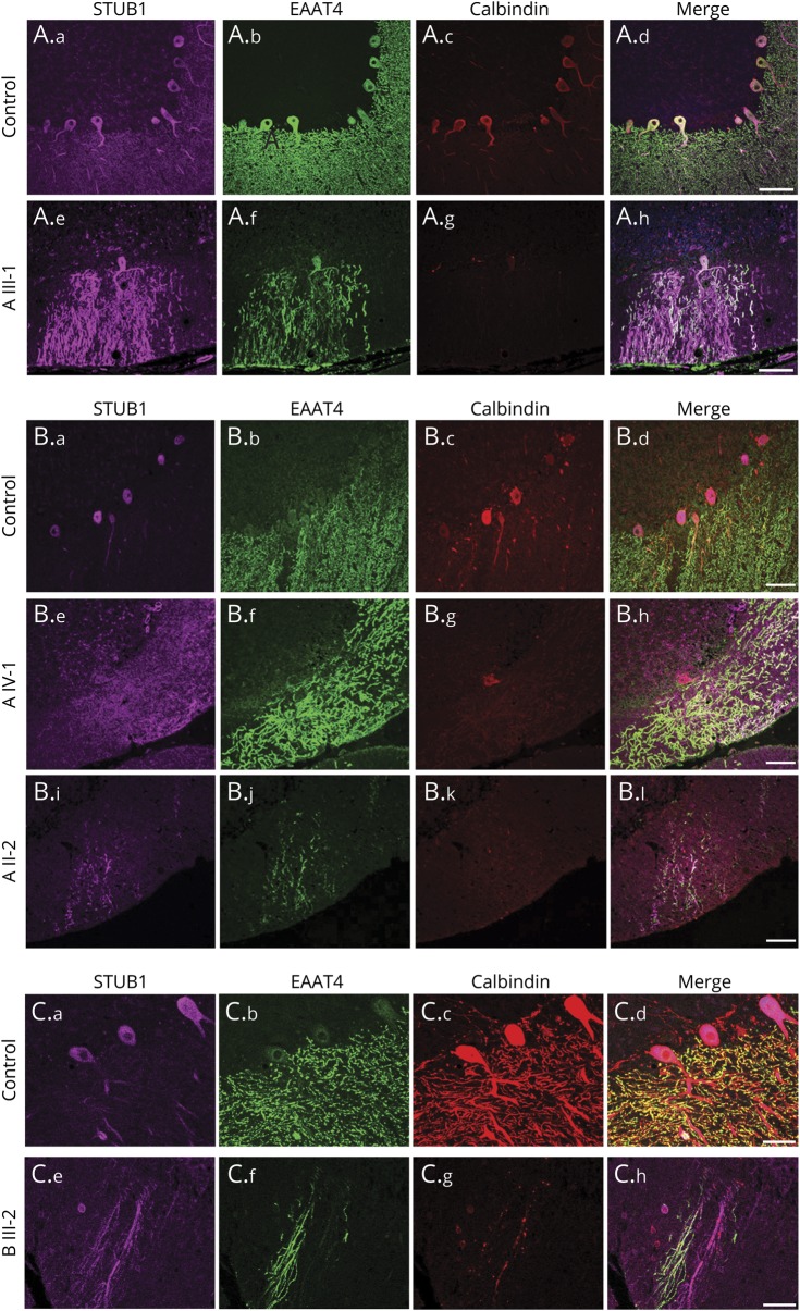
Figure 5. Loss of polarized STUB1 expression in Purkinje cells in patients with STUB1-cerebellar ataxia.
Immunostaining was performed with antibodies to STUB1 (pseudocolored purple), EAAT4 (green), and calbindin (red), and all scanning acquisition and postprocessing parameters were carried out identically among the cases about the controls in each panel. In 3 different normal control individuals (A.a–A.d, B.a–B.d, C.a–C.d), STUB1 immunoreactivity was uniformly localized primarily in PC cell bodies and proximal dendrites, with little expression in distal PC dendritic arbors. (A and B) Family A with STUB1-Ile53Thr. Loss of STUB1 polarization results in aberrant expression in distal PC dendritic arbors and cell bodies in patients III-1 (A.e–A.h), IV-1 (B.e–B.h), and II-2 (B.i–B.l). Scale bars = 100 μm. (C) Family B with STUB1-Phe37Leu. In individual III-2, STUB1 was aberrantly expressed in PC distal arbors (C.e–C.h). Scale bars = 50 μm.

