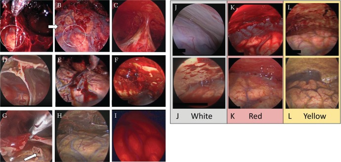
Fig. 1.
Endoscopic findings in the chronic subdural hematoma cavity. A soft hematoma on the inner membrane was carefully suctioned by a suction tube to observe the hematoma cavity in detail (A and B). The circles in A and B show the same parts. Trabecula (C), septum (D), clots adhering to the inner membrane (E), clots adhering to the outer membrane (F), an apparent tear of the arachnoid mater (G), a yellow inner membrane (H), and a red membrane (I). The septum was endoscopically divided using scissors (G). The color groups of the outer membrane (J–L). The white group presents an evenly white outer membrane without capillaries and with/without patchy clots like erythema and purpura (J). The red group presents with an evenly red or pink outer membrane with/without hemorrhage or hematoma adhering to the outer membrane (K). The yellow group presents as yellow or orange but not evenly red or white outer membrane with capillaries developing apparently (L).

