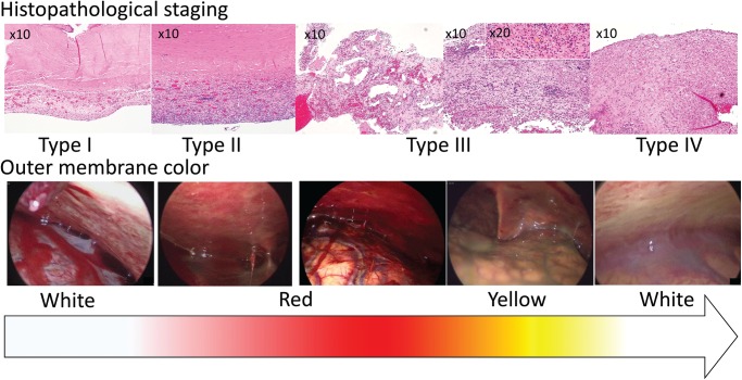
Fig. 2.
The histopathological specimens and endoscopic findings of the same patients were lined up in order of histopathological staging. As the histopathological staging changes from type I to IV (upper row), the outer membrane color changes from white to red to yellow to white in order (lower row).

