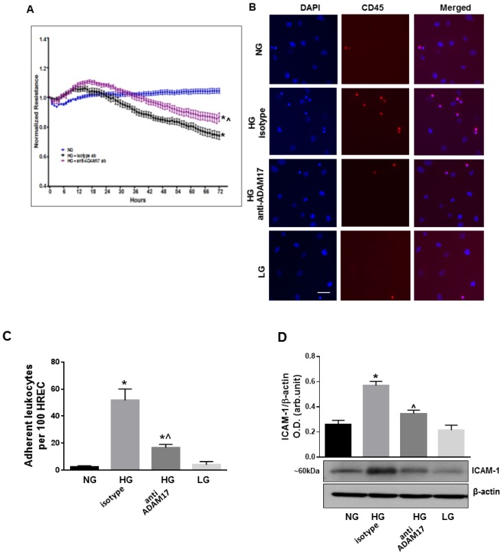
Figure 7.
Effects of ADAM17 blocking antibody on high glucose-induced endothelial cell permeability, leukocyte adhesion and ICAM-1 expression. (A) TER in HREC monolayers treated with HG in the presence of ADAM17 blocking antibody or control isotype antibody using ECIS. Data presented as mean ± SE. * p < 0.05 NG vs. HG. ^ p < 0.05 HG + isotype vs. HG + anti-ADAM17 antibody. n = 3. (B) Representative images of leukocytes adherent to HREC exposed to HG in the presence of 0.5 µM ADAM17 blocking antibody D1(A12) or control isotype antibody. Scale bar, 50µm. (C) Quantitative analysis of leukocyte adhesion. Data are expressed as a number of adherent cells per 100 HREC. * p < 0.05 NG vs. HG. ^ p < 0.05 HG + isotype vs. HG + anti-ADAM17 antibody. n = 3. (D) Representative blots and densitometric analysis of ICAM-1 expression in HREC treated with HG in the presence of ADAM17 blocking antibody or control isotype antibody. Data presented as mean ± SE. * p < 0.05 NG vs. HG + isotype antibody. ^ p < 0.05 HG + isotype vs. HG + anti-ADAM17 antibody. n = 4.

