Figure 4.
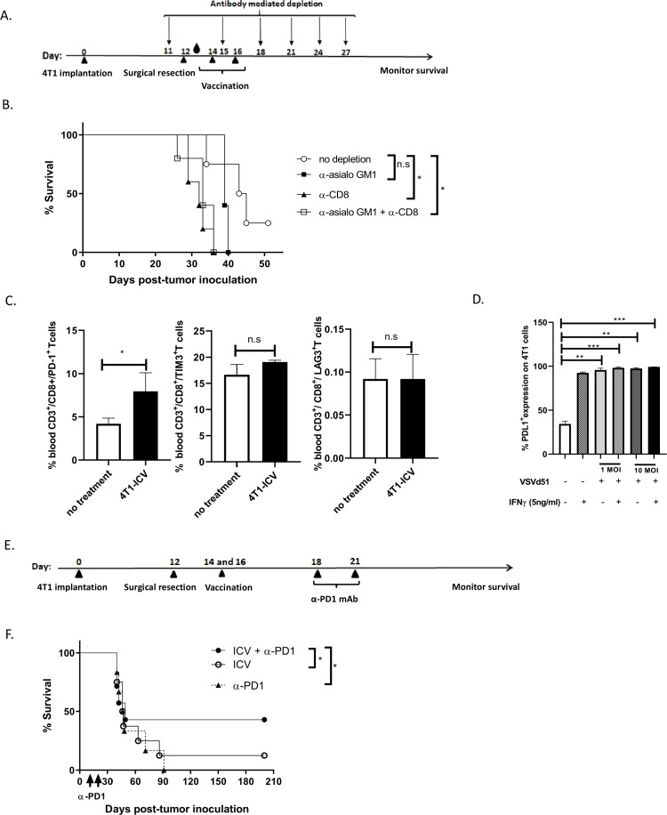
CD8+ cytotoxic T cells are critical for infected cell vaccine (ICV) efficacy and combination treatment with anti-PD1 checkpoint inhibitor improves survival in BALB/c-4T1 model. (A) Timeline of immune cell depletion in the BALB/c-4T1 in vivo model. One day before surgical resection, NK cells, CD8+ T cells and NK+CD8+ T cells were depleted using antibodies to GM1, CD8 and GM1+CD8, respectively, and continued every 3–4 days for a total of 6 doses. On days 14 and 16, mice received 2 doses of ICV. Blood droplet denotes verification of in vivo depletion by flow cytometry. (B) Kaplan-Meier survival analysis of BALB/c mice bearing intramammary 4T1 tumors and receiving ICV and antibody depletion. n=10–12 mice/group. *p<0.05; n.s., not significant, log-rank test. (C) Single cell suspensions from the peripheral blood of mice following indicated treatments were stained with exhaustion markers on CD8+ T cells (PD1, Tim3, LAG3) and analyzed by flow cytometry. All data are representative of three similar experiments where n=3–5 mice/treatment. *p<0.05; n.s., no significance. (D) Cell surface staining of PD-L1 on 4T1 cells following infection with VSVd51 in the presence or absence of IFNγ and analyzed by flow cytometry. (E) Timeline of combination therapy ICV+αPD1 in the BALB/c-4T1 in vivo model. Two days after vaccination, mice received 2 doses of anti-PD1 intraperitoneally 3 days apart. (F) Kaplan-Meier survival analysis of BALB/c mice bearing intramammary 4T1 tumors and receiving ICV and anti-PD-1. n=10–12 mice/group. *p<0.05; n.s., not significant, log-rank test.

