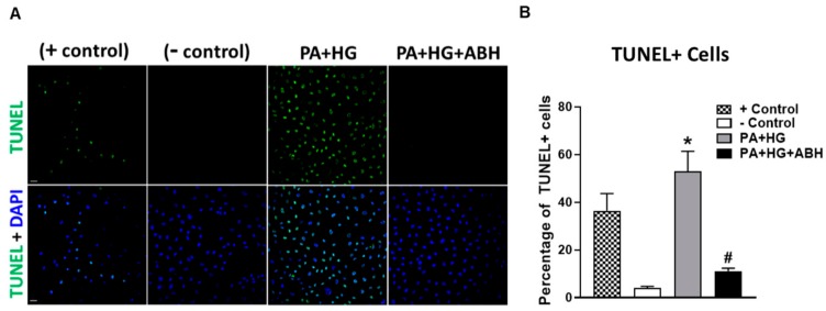
Figure 9.
Representative terminal deoxynucleotidyl transferase dUTP nick end labeling (TUNEL) photomicrographs at 20× with quantitation (A,B) showing increased percentage of TUNEL positive cells in bovine retinal endothelial cells (BRECs) treated for 72 h with PA+HG (scale bar = 100 µm, n = 5–7 per group). Cell nuclei are labeled with DAPI (blue) and TUNEL positive cells fluoresced green. Data are presented as mean ± SEM (standard error of the mean). * p < 0.05 compared to control (BSA-treated) group, # p < 0.05 compared to PA+HG-treated group. Scale bar = 50 μm.

