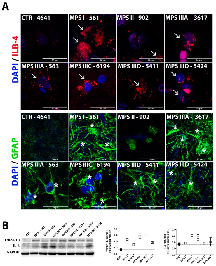
Figure 3.
Astromicrogliosis in brain cortex tissues of human MPS patients is indicative of neuroinflammation. (A) Confocal microscopy images of brain cortex tissues of MPS I (561), MPS II (902), MPSIIIA (3617 and 563), MPS IIIC (6194), and MPS IIID (5411 and 5424) patients and a representative control (4641) stained with isolectin beta-4 (ILB-4,red) and antibodies against GFAP (green), markers for activated microglia and astrocytes, respectively. The fixed brain tissues of the MPS II patient HBCB1801OC were not sufficiently preserved to perform cryosectioning and conduct immunofluorescent analysis. DAPI (blue) was used as the nuclear counterstain. Activated microglia are marked with arrowheads and astrocytes are marked with asterisks; scale bar: 20 µm. (B) Western blotting analysis showing increased protein expression of the proinflammatory cytokines (tumor necrosis factor superfamily member 10 (TNFSF10) and interleukin 6 (IL-6) in the brain cortex protein extracts from MPS patients and combined controls (662, 754, 1266, 4641, 5287, 5813, and 5977). Glyceraldehyde 3-phosphate dehydrogenase (GAPDH) was used as a loading control. Data are expressed as the mean ± s.e.m.

