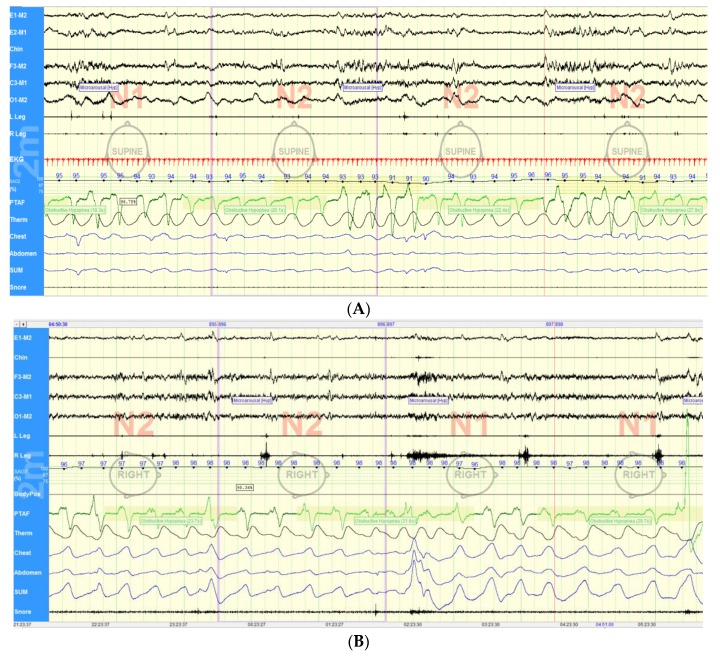
Figure 2.
Patterns of upper airway obstruction in Parkinson’s disease. (A) Obstructive hypopneas associated with microarousals and oxygen desaturation in a patient with Parkinson’s disease and obstructive sleep apnea. (B) Upper airway instability in a patient with Parkinson’s disease resulting in obstructive breathing. PTAF, pressure transducer airflow and Therm, thermistance. Chest and abdomen refer to the respective position of the bands used to detect respiratory efforts and SUM correspond to the sum of the chest and abdominal bands’ signal.

