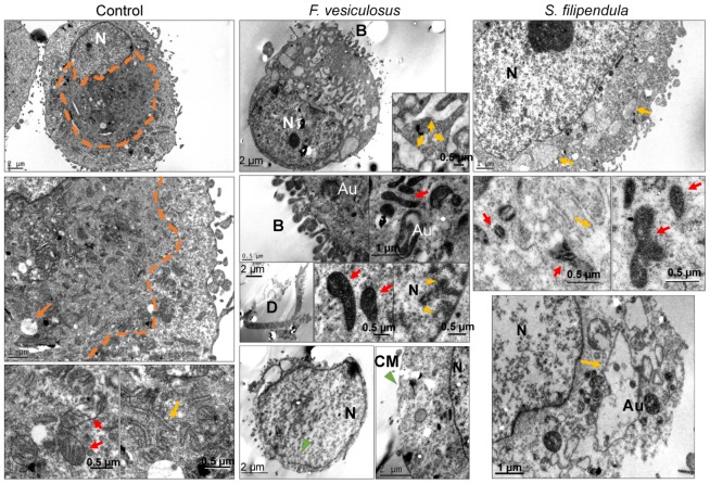
Figure 3.
TEM of MG63 cells treated with 100 μg/mL of crude fucoidan from F. vesiculosus or S. filipendula (at least 10–15 cells analysed per condition). Red arrows—mitochondria, orange arrows—vesicles or vacuoles, orange dashed line—perinuclear region rich in organelles in control cells, N—nucleus, B—blebbing in the cell membrane, D—cellular debris, yellow arrow heads—chromatin condensation and marginalisation, green arrow heads—membrane nicks, yellow arrows—endoplasmic reticulum at higher magnification.

