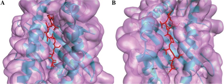
Figure 3:
Docking predicts the interaction of predicted epitopes to MHC class I molecule, HLA-A*02:03 and HLA-B*35:01.
Binding of “ALQTGITLV” to the interacting grooves of the generated structure of HLA-A*02:03 (binding energy: −8.4 Kcal/mol). Binding of “VPSSSTPL” to the binding grooves of the retrieved structure of HLA-B*35:01. (Binding energy: −8.5 Kcal/mol) The blue colored portion and green portion in both figure (A and B) represent HLA-A*02:03 molecule, ALQTGITLVL epitope and HLA-B*35:01 molecules and VPSSSTPL epitope, respectively.

