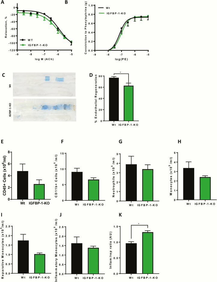
Figure 4.
IGFBP-1 knockdown and endothelial function. (A) Relaxation responses to acetylcholine (Ach), expressed as percent reversal of phenylephrine induced contraction was the same between the genotypes. (B) Constrictor responses to PE was the same between the genotypes (N = 3–4). C: Representative in situ Evans blue staining 5 days after vascular injury (blue staining indicates denuded endothelium) in WT and IGFBP-1-KO mice (magnification ×20) (Sham bottom). (D) Endothelial regeneration 5 days after vascular injury is reduced in IGFBP-1-KO mice when compared to litter mate controls (IGFBP-1-KO 62.82 ± 4.5 V Wt 76.9 ± 2.1) (N = 4–6 per group). (E–J) There is no difference in CD45+ cells (IGFBP-1-KO 2.59 ± 0.7 V Wt 4.78 ± 1.2 x10⁶/mL), CD11b+ cells (IGFBP-1-KO 6.6 ± 0.56 V Wt 9.08 ± 1.16 × 10⁵/mL), neutrophil numbers (IGFBP-1-KO 3.3 ± 0.46 V Wt 3.8 ± 0.86 x10⁵/mL), monocyte cells (IGFBP-1-KO 2.4 ± 0.14 V Wt 3.37 ± 0.06 × 10⁶/mL), reparative monocytes (IGFBP-1-KO 1.023 ± 0.059 V Wt 1.75 ± 0.32 × 10⁶/mL) or inflammatory monocytes (IGFBP-1-KO 1.38 ± 0.08 V Wt 1.62 ± 0.33 × 10⁶/mL) in IGFBP-1-KO mice when compared with controls. (K) There is a significant skewing of inflammatory to reparative monocytes IGFBP-1-KO mice when compared with controls (IGFBP-1-KO 1.32 ± 0.04 V Wt 0.96 ± 0.05) (n = 5). Data are presented as mean ± SEM. (*P ≤ .05).

