
Figure 4.
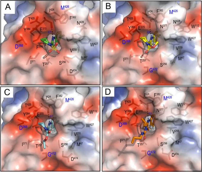
Protein–analog structures with LM/HT gp120CRF01_AE core: A, 6l (PDB code: 6ONV); B, 16a (PDB code: 6ONE); C, 16b (PDB code: 6ONF); D, 17d (PDB code: 6ONVH). Structures were aligned based on gp120, and the electrostatic potential is displayed over the gp120 molecular surface colored red for negative, blue for positive, and white for apolar. Analogs are shown in a ball–stick representation, and residues lining the analogs binding cavities (buried at the interface as calculated by PISA software) are shown as sticks. There is one direct H-bond at the contact interface of each analog formed between Thr425 O of gp120 and amide N of the compound. Key conserved residues in the Phe43 pocket: Asp368, Gly472, and Met426 are labeled in blue.