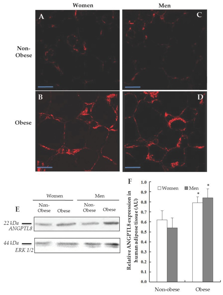
Figure 2.
Detection of ANGPTL8 in human visceral adipose tissue (VAT). Representative images from the immunofluorescence reaction for positive ANGPTL8 (in red) in adipose cells of patients, (A, non-obese women, n = 4), (B, obese women, n = 4), (C, non-obese men, n = 4), (D, obese men, n = 4). Scale bar: 60 μm. (E) Western blotting analysing the expression of ANGPTL8 in human VAT (n = 6), ERK1/2 was used to confirm equal loading of samples. (F) Plot showing the relative ANGPTL8 expression as arbitrary units (AUs).

