Figure 1.
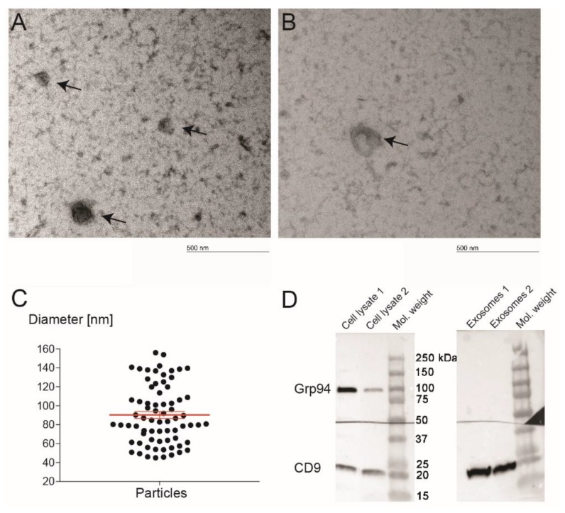
The characterization of exosomes derived from dental pulp. TEM was used to study the structure and the size of exosomes (A,B). Image of an extracted exosome showing the typical lipid bilayer structure (B). The scale bar represents 500 nm. Microscope magnification was 66,000×. It was observed that the diameter of isolated particles was 90 ± 30 nm (C). Western blot results showed that the key exosomal membrane protein CD9 was positive for exosomes, while the cell protein Grp94 was negative for exosomes (D). The analysis verified that the isolated vesicles were indeed exosomes.

